Sundar Spine

For Appointments, Call

+(91) 9080 9680 66

Fracture Surgery

Plate & Screw Fixation

Fracture fixation using Plates & Screws

Proximal Femoral Nailing

Rod fixation for Hip Fractures

External Fixation

By Pins and rods outside the body

Intramedullary Nailing

Long rod used for Fracture fixations

K-Wire Fixation

Thin wire Fracture fixation

Plate & Screw Fixation

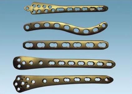
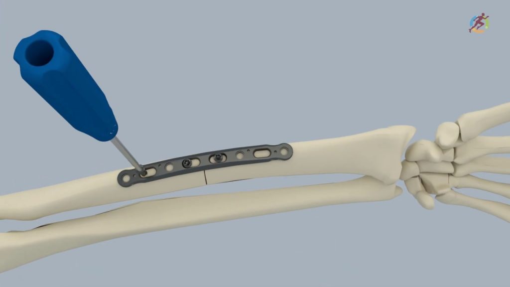
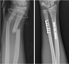
Plate and screw fixation of fracture is one of the most common orthopaedic intervention for fracture management. The procedure involves reducing the displaced fracture fragments and fixing it rigidly by a long linear metal implant (plate) to the bone. Screws are used to facilitate this fixation. 

When is this surgery performed?

This is one of the most common and successful orthopaedic surgery, used when:

  • The fracture is displaced
  • Fracture cannot heal properly with immobilization alone
  • Multiple fracture fragments
  • Weight/stress bearing bone
  • Intra-articular fracture
How is the surgery performed?

The surgery is performed by the concept of Open reduction internal fixation (ORIF). The steps involved in any plate fixation are as follows:

  • The surgery is done by General, Spinal or regional anasthesis depending on the site of fracture and surgeons preference.
  • An incision is made over the fractured bone (based on years of research done to reduce the number of muscle split and safely avoid any nerves and blood vessels)
  • The fracture site is approached by retracting the overlying muscles and structures.
  • The fracture is reduced with the help of special instruments and reduction is confirmed both visibly and by C-arm (xray machine).
  • A plate of appropriate length is selected and placed over the bone and fixed on either end using multiple screws (number of screws deemed necessary by the surgeon).
  • The reducion instruments are removed and the fracture stability is assessed by moving the joints above and below.
  • Once the fixation is confirmed to be stable the wound is closed and dressing applied.
  • Sometimes a plaster slab may be applied in certain fractures to reduce post-operative edema, added stability and faster recovery.

Lifting heavy weights is avoided for few days in upperlimb fracture fixation. Weight-bearing is avoided for few days in lower limb fracture fixation.

Recovery time:
The recovery time in fracture fixation is higly dependant on the bone fractured, type and severity of fracture, stability of surgical fixation and the bone quality. It is advised to follow the doctors advise regarding walking, hospital stay, resuming work, driving car/bike, other activities and rehabilitation.

Intramedullary Nailing

Intramedullary nailing (IM Nailing) is a technique of fracture fixation commonly used in long bones – thigh bone (femur), Leg bone (tibia) and the arm bone (humerus). This technique has is mainly prefered for weight-bearing bones as they give more stability and support early walking after surgery.

When is this surgery performed?

This technique can be done only in very specific type of fractures:

  • Shaft (middle) and proximal fracture of the thigh bone (femur)
  • Shaft (middle) fracture of the leg bone (tibia)
  • Shaft (middle) fracture of the arm bone (humerus)

Advantages of this procedure:

  • Early weight bearing
  • Smaller incision (compared to plating)
  • Incision not over the fracture site – faster healing
  • Lesser blood loss
  • More stable
  • Faster bone union

How is the surgery performed?

The surgery is performed by the concept of Closed reduction and internal fixation (CRIF). The steps involved in any intramedullary nailing are as follows:

  • The surgery is done by Spinal anasthesia (lower limb) and general or regional anasthesia (upper limb).
  • An incision is made over one end of the bone near the joint.
  • The bone is entered with a sharp instrument and the hollow middle (medullary cavity) of the bone is approached.
  • A guide wire is inserted through the hole which is passed to the full length of the bone crossing the fracture site.
  • Certain manipulation is required to pass the guide wire across the fracture site. (confirmed by C-arm)
  • The medullary cavity is reamed (widened) to accomodate the implant – nail.
  • The implant – Intramedullary Nail is introduced through this canal and fixed by screws both far above and below the fracture site.
Recovery time:
The recovery time in fracture fixation is higly dependant on the bone fractured, type and severity of fracture, stability of surgical fixation and the bone quality. It is advised to follow the doctors advise regarding walking, hospital stay, resuming work, driving car/bike, other activities and rehabilitation.
Nailing-scaled
nail-xray-scaled

Proximal Femoral Nailing

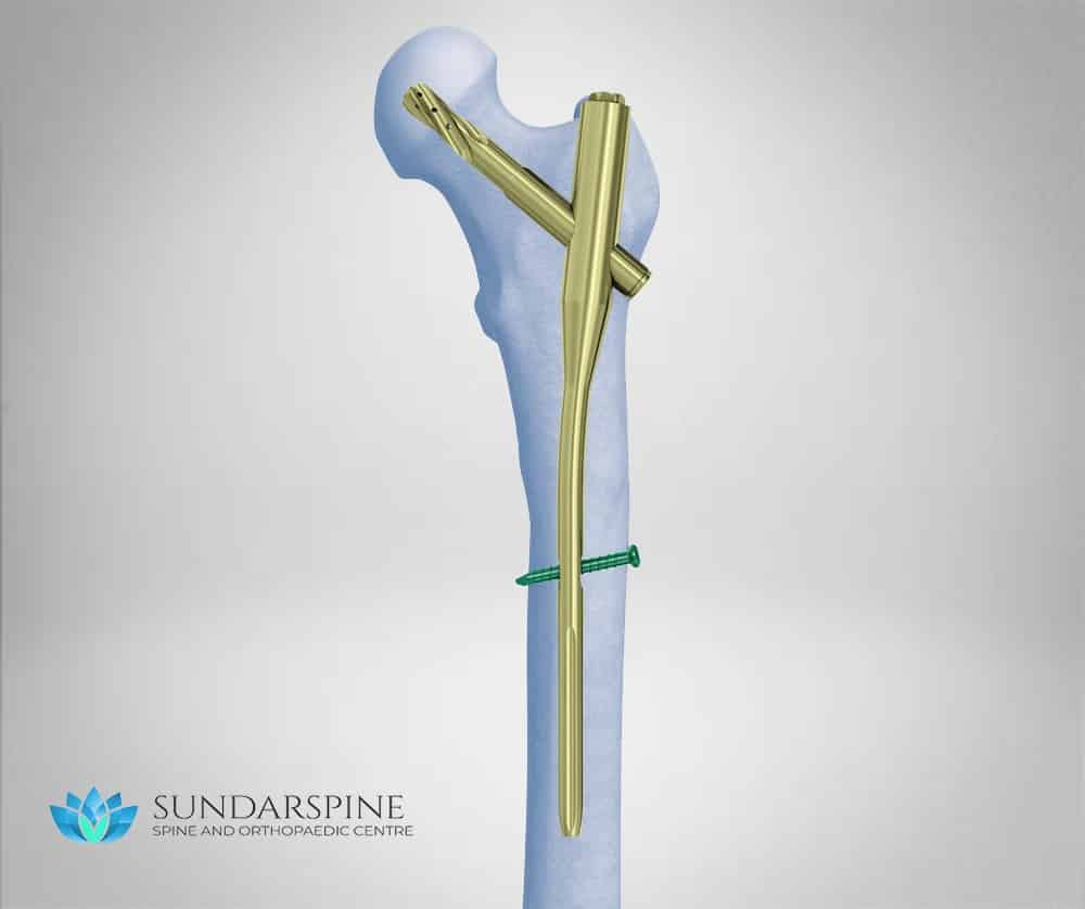
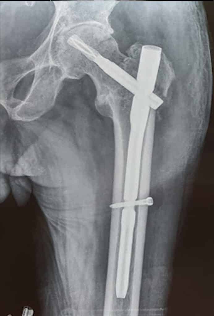
Proximal Femoral nailing is a technique of fracture fixation that is used to treat hip fractures (Proximal femur). Hip fractures are the most common type of fracture especially in the elderly. 

When is this surgery performed?

This technique can be done only in very specific type of fractures:

  • Hip fracture (intertrochanteric)
  • Upper thigh fracture (Subtrochanteric)

Advantages of this procedure:

  • Early weight bearing
  • Smaller incision (compared to plating)
  • Incision not over the fracture site – faster healing
  • Lesser blood loss
  • More stable
  • Faster bone union
How is the surgery performed?

The surgery is performed by the concept of Closed reduction and internal fixation (CRIF). The steps involved  are as follows:

  • The surgery is generally done by Spinal anasthesia.
  • The fracture is reduced by manipulation on a fracture table before incision.
  • An 8-10cm incision is made over the uppermost end of the thigh where the thigh bone (femur) starts.
  • The bone is entered with a sharp instrument and the hollow middle (medullary cavity) of the bone is approached.
  • A guide wire is inserted through the hole which is passed to the full length of the bone crossing the fracture site.
  • The medullary cavity is reamed (widened) to accomodate the implant – proximal femoral nail.
  • The implant may be a short nail (intertrochanteric fracture) or a long nail (subtrochanteric fracture).
  • The nail is fixed by using a special blade/screw in the upper side and normal screw on the lower side though seperate small incisions.
Recovery time:
Duration of surgery: 1 to 2 hours
Walking started: 24-48 hours (level of weight bearing depends upon fracture pattern and fixation stability)
Hospital stay: 2-4 days
Resume work: 8 weeks
Brace : Nil
Driving (Car, Bike): 12 weeks
Strenous Activity (bending down, lifting weigths): 3 months 

K-Wire Fixation

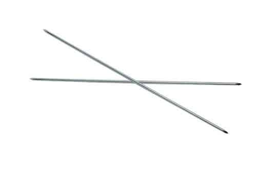
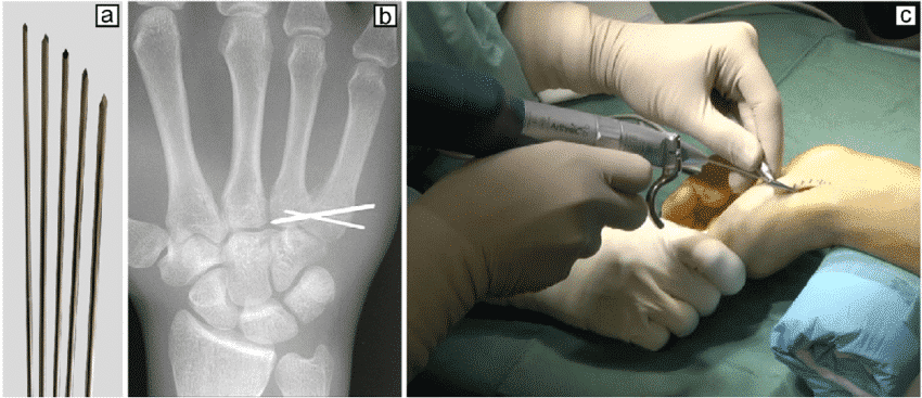
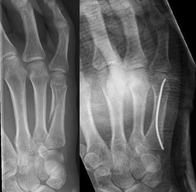
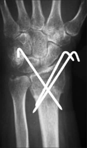
K-wire is a thin metallic pin/wire like structure that is used to fix or stabilize a fracture. They are normally used to fix fractures of the small bones (hands or foot) or larger bones in children. They do not have inherent stability, hence are usually used along with splints or braces till fracture stability.

How the surgery is performed?

  • The procedure is usually performed under light sedation or regional block depending on the age and location of fracture.
  • The fracture is reduced by manipulation under C-arm
  • The k-wire of appropriate diameter is introduced with the help of a drill. 
  • The wire is usually engaged into the opposite bone across the fracture holding both the fragments in reduced position.
  • Usually more than one k-wire might be required to fix fragments in both planes.
  • The other end of k-wire is usually left outside or can be buried inside the skin with sutures.
  • The procedure is usually supplemented by a splint or a brace.

The k-wire necessitates removal after 3-6 weeks. The exposed k-wire can be removed in outpatient procedure room whereas the buried k-wire require local/regional anasthesia and hence might be done as an operative room procedure. 

Advantages of K-wire:
Holds the fracture fragments in reduced position till the fracture heals. Multiple k-wires increase the stability of the fracture fixation.

Recovery time:

Duration of surgery: 1-2 hours 

Walking started: Variable

Hospital stay: 1-2 days 

Resume work: After k-wire removal
Brace (Splints): 3-6 weeks

Driving (Car, Bike): After k-wire removal

Strenuous Activity (bending down, lifting weights): Variable

External Fixator

  • External Fixator is a technique of holding the fracture fragments by a pin connected to a rod externally, until the skin and soft tissue heals.

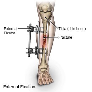
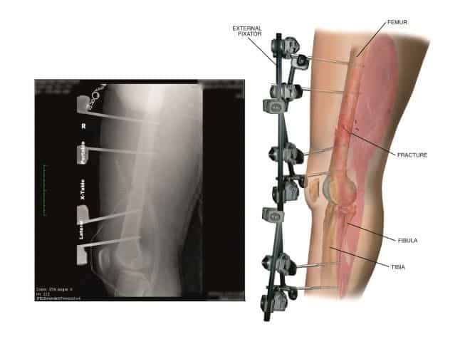
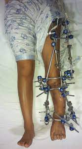
This procedure is usually done in open fracture or in severe multiple injuries to give adequate time for healing before the main final fixation.

When is this Surgery performed?

  • In severe open fracture
  • Multiple long bone or pelvic fracture
  • Complex fracture near joints (knee, wrist or ankle)
  • Fracture with associated life threatening injuries.
  • As a temporary stabilization till the patient is fit for the main procedure.
How the surgery is performed?
  • The surgery can be performed under general, regional or spinal anasthesia.
  • The fracture is reduced under C-arm guidance and multiple pins are placed above and below the fracture site.
  • All the pins are connected by a rod which holds the fracture in relative stability.

Advantages of External fixator:

This procedure is usually done as a temporary stabilization till the would and the patient are stable for further procedure. The initial 3 days after a high velocity is a risk period for both the wound (increased swelling, infection) and the patient (risk to life). Hence major procedure such as final fixation are avoided during this period.

Recovery time:
Duration of surgery: 1 to 2 hours 
Walking started: Non-weight bearing
Using Restroom: Variable
Hospital stay: Variable
Resume work: After final fixation
Brace: Nil
Driving (Car, Bike): After final fixation
Strenous Activity (bending down, lifting weigths): After final fixation 
493px-External_fixator_xray

Vertebroplasty

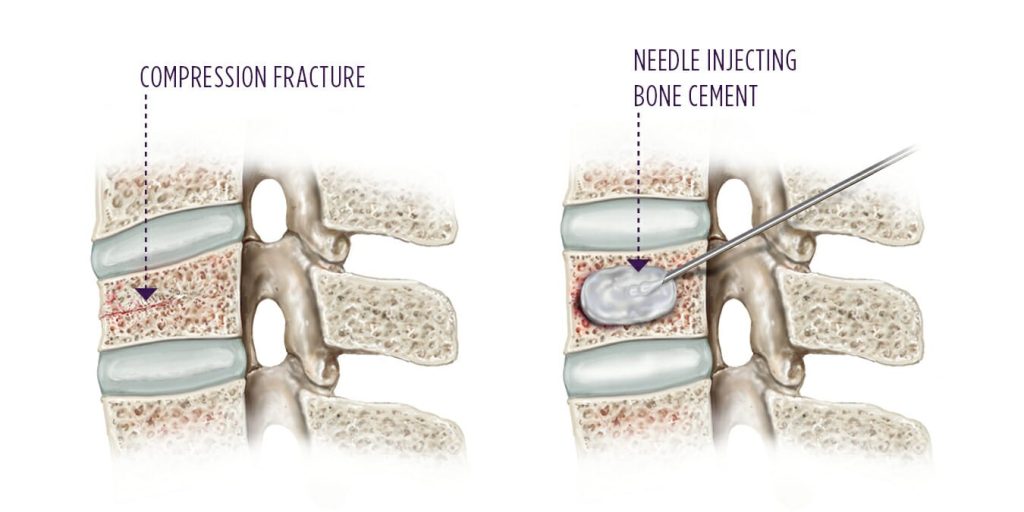
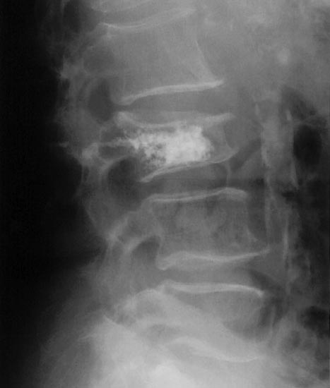
Vertebroplasty is an outpatient procedure for stabilizing compression fractures in the spine. Bone cement is injected into back bones (vertebrae) that have cracked or broken, often because of osteoporosis. The cement hardens, stabilizing the fractures and supporting your spine.

For people with severe, disabling pain caused by a compression fracture, vertebroplasty can relieve pain, increase mobility and reduce the use of pain medication.

When the surgery is performed? 

Osteoporotic fracture (more than 60% collapse)

How the surgery is performed?
  • The procedure is usually performed in local anasthesia with mild sedation.
  • The vertebral level is marked using a C-arm (xray machine).
  • Two bone biopsy needles are inserted on either side of the midline through the pedicles of the involed vertebra till it reached the far side of the vertebra.
  • Bone cement is injected into both the needles and systematically withdrawn such that much of the vertebra is covered by the cement. 
  • Care is take that bone cement does not spill out of focus area especially posteriorly into the spinal canal.
  • The needles are withdrawn and the needle entry site are closed by bandages without any sutures.

Recovery time:

Duration of procedure: 1 hours
Walking started: 2-4 hours
Hospital stay: 0-1 days
Resume work: 1 weeks
Brace (Cervical collar): 1 week
Driving (Car, Bike): 2 weeks
Strenous Activity (bending down, lifting weigths): 1-2 months 

Kyphoplasty

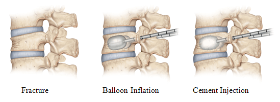
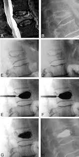
Kyphoplasty is similar to vertebroplasty, but uses special balloons to create spaces within the vertebra that are then filled with bone cement. Kyphoplasty can correct spinal deformity and restore lost height

When the surgery is performed? 

Tuberculosis spine, kyphosis deformity, vertebral collapse, tumour spine

How the surgery is performed?
  • The procedure is usually performed in local anasthesia with mild sedation.
  • The vertebral level is marked using a C-arm (xray machine).
  • A canula is inserted through the pedicle into the vertebral body.
  • The canula is inflated like a balloon which creates a cavity in the vertebral body simultaneously correcting the kyphosis.
  • Bone cement is injected through the canula such that the cavity is filled.
  • Small bandage is placed to cover the entry site.

Recovery time:

Duration of surgery: 1 hour
Walking started: 6-12 hours
Hospital stay: 1-2 days
Resume work: 3-4 weeks
Brace (Cervical collar): 4-8 week
Driving (Car, Bike): 8 weeks
Strenous Activity (bending down, lifting weigths): 2-3 months  

Scoliosis Surgery

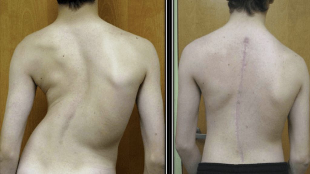
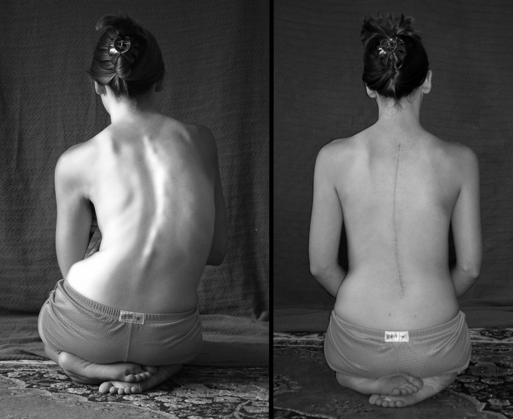
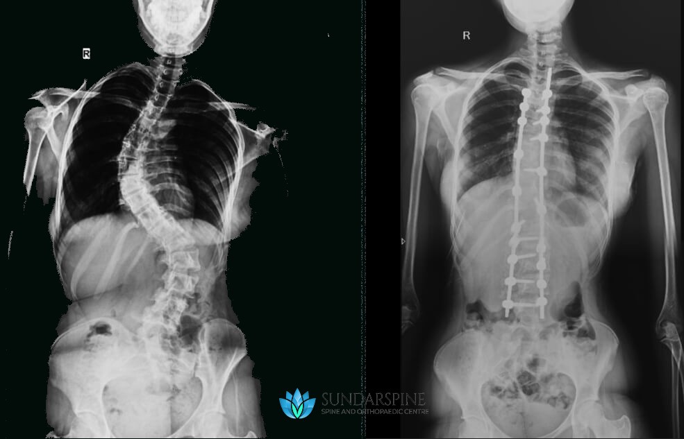
The operation for scoliosis correction is a spinal fusion. The basic idea is to realign and fuse together the curved vertebrae so that they heal into a single, solid bone.

With the tools and technology available today, scoliosis surgeons are able to improve curves significantly.

When the surgery is performed? 

 The decision to go ahead with scoliosis correction surgery depends on the age of the patient, severity of deformity, secondary cause, curve type and  curve progression. However, most scoliosis surgeons agree that children who have very severe curves (45 to 50 degrees and higher) will need surgery to lessen the curve and prevent it from getting worse.

Advantages of Scoliosis Correction:

  1. Stop and reverse curve progression
  2. Prevent respiratory and cardiac complications
  3. Cosmetic improvement
  4. Prevent development of backpain
  5. Improve the lifestyle of the patient
How the surgery is performed?
  • The surgeon and team perform a detailed evaluation of the patient and the curve for surgical planning. This involves multiple xrays in various bending and stretching positions.
  • The procedure is performed under general anasthesia usually with Neuro monitoring.
  • A long linear midline incision is placed on the back over the levels to be corrected.
  • The muscles are systematically retracted and the pedicle screws (in levels preplanned) are applied.
  • The lamina bone is removed and soft tissues are released which will make the spine flexible. 
  • A pre-bent rod is placed and methodically fixed and rotated to recreate the spine alignment. Bone grafts are placed on the sides for added fusion support.
  • The wound is usually long involving multiple stitches and a big dressing.

In severe curves, an anterior surgery might also be necessary and will be performed in the same day or as a 2-stage (alternate day) procedure.

Recovery time:

Duration of surgery: 4-10 hours (Depends on the number of levels)
Walking started: 24-48 hours
Hospital stay: 4-8 days
Resume work: 12 weeks
Brace (Cervical collar): 12 week
Driving (Car, Bike): 12 weeks
Strenous Activity (bending down, lifting weigths): 6 months 

Endoscopic Surgery

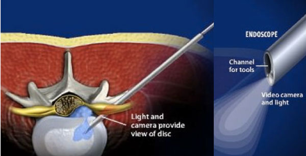
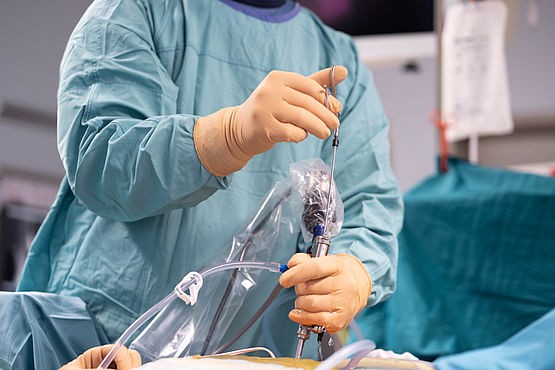
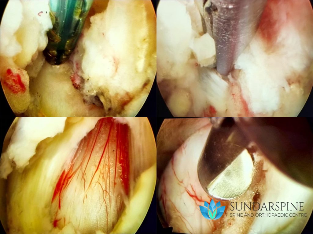
Endoscopic spine surgery is one of the latest advancement in the field of medicine. With the use of a specialized micro-camera and modern crafted instruments, many spine conditions can be treated with a portal (incision) less than 8mm in size.

When the surgery is performed? 

Endoscopic spine surgery is mainly performed for Lumbar disc herniation without any significant bone spur or instability.

Advantages of Endoscopic Surgery:

  1. Small incision (less than 1 cm)
  2. Very minimal post-operative pain
  3. Same day discharge
  4. No blood loss
  5. Faster return to work
  6. No surgical scar
How the surgery is performed?
  • The procedure can be performed under general anasthesia or with local and light sedation.
  • The level of surgery is marked with the help of C-arm (xray) precisely before starting the procedure.
  • A 8mm incision is made on the premarked area at the back (interlaminar approach) or at the sides (transforaminal approach)
  • A dilator is used to clear a path followed by a canula and camera.
  • Series of c-arm shots will be taken to confirm the camera is in correct level and position.
  • A path to the diseased disc may be cleared and all the offending fragments of the disc are removed.
  • Wound closed by a single suture. 

Recovery time:

Duration of surgery: 1-2 hours (Depends on the number of levels)
Walking started: 4-8 hours
Hospital stay: 0-1 day
Resume work: 2 weeks
Brace (Cervical collar): 1 week
Driving (Car, Bike): 3 weeks
Strenous Activity (bending down, lifting weigths): 3 weeks